At Rawlings, we believe your eyesight deserves more than just a basic check-up. That’s why every one of our branches is equipped with cutting-edge Optical Coherence Tomography (OCT) — the gold standard in retinal imaging.
We also continue to offer retinal photography, giving you both modern insight and visual records of your eye health.
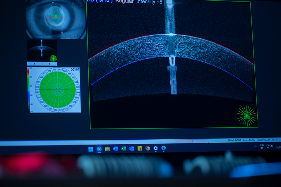
Optical Coherence Tomography (OCT) is a state-of-the-art, non-invasive imaging technique that allows us to see deep beneath the surface of your retina in exceptional detail. Unlike standard retinal photography, which captures a surface-level snapshot of the back of your eye, OCT provides a high-resolution, 3D cross-sectional view – enabling early detection of potential issues long before symptoms appear.
Think of retinal photography as the postcard – and OCT as the full documentary. Both have value, but only one tells the whole story.
OCT scans are vital for early detection and monitoring of a wide range of serious eye conditions, including:
We securely store your scans to allow direct comparisons over time – helping your Optometrist detect even the most subtle changes in your eye health, visit by visit.
Your OCT scan takes just a few seconds to complete during your regular eye exam when pre-booked. It’s:
While not covered by NHS eye tests, OCT is available as a premium add-on for a small additional fee.
We also allocate extra time with your Optometrist to ensure your results are fully explained and any next steps are clearly understood.
Members of our Rawlings Vision Plan enjoy OCT screening as part of their plan. Not yet a member? You can join on the day of your appointment and still benefit from full OCT screening at no extra cost.
Take control of your eye health with advanced retinal imaging from Rawlings. Whether you’re new to OCT or already enjoy the benefits, we’re here to ensure you receive the most thorough care available.
To pre-book your OCT scan or find out more about pricing, visit our fees page or speak to a member of our team when you book your appointment.
With practices across Hampshire and Surrey, expert eye and hearing care is never far away. Enter your postcode below to find the nearest Rawlings branch.